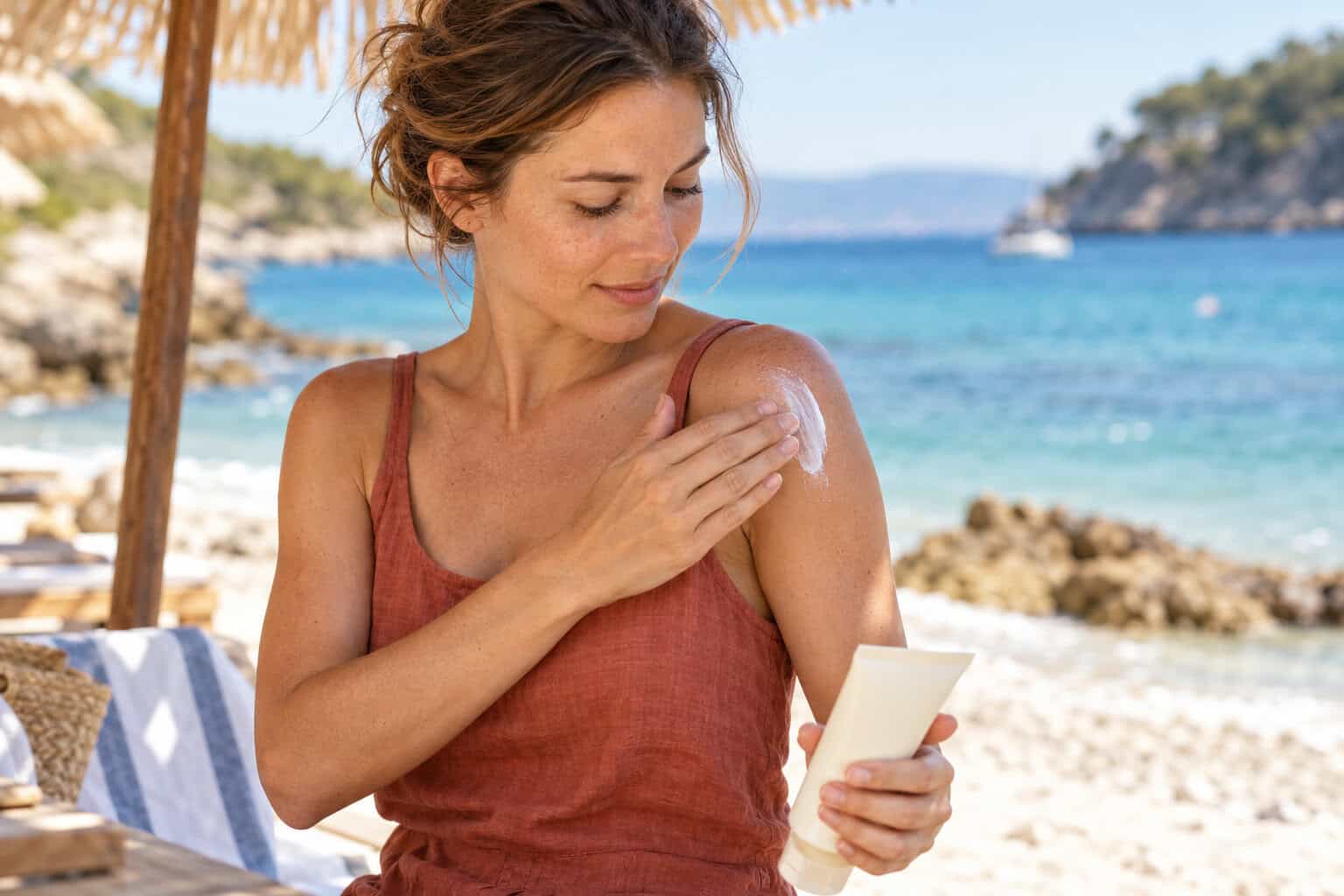
Every stretch of strong summer sun sets off a quiet demolition job inside your skin. Ultraviolet B light, the shorter wavelength behind sunburn, switches on enzymes that chop up collagen, the protein scaffolding that keeps skin firm. Over decades the damage adds up to wrinkles, sagging and uneven pigment, which dermatologists call photoaging. A new skin photoaging study now asks whether hemp seed nanoparticles, microscopic bubble-like packages pulled out of ordinary hulled hemp seeds, can tip that balance back toward repair.
The work, by Hannah S. Park and Sehyun Shin of the Engineering Research Center for Biofluid Biopsy and the School of Mechanical Engineering at Korea University in Seoul, was published at the end of September 2026 in the peer-reviewed journal Molecules (DOI: 10.3390/molecules31193455). It appeared in a special issue on natural cosmetic ingredients, and the full text is open access. One point matters before anything else: every result in the paper comes from cells grown in lab dishes or from test-tube assays. No volunteer put anything on their face.
Key Takeaways
Korean researchers isolated vesicle-like nanoparticles from dehulled hemp seeds, mostly between 100 and 200 nanometres across, and tested them on human and mouse cells. In human skin fibroblasts, the cells that make collagen, the particles raised production of procollagen type I by about 45% and pushed down the UVB-triggered surge of MMP-1, an enzyme that breaks collagen down. They also helped skin cells close an artificial scratch faster and cut pigment production in mouse pigment cells. The picture on inflammation was mixed rather than uniformly calming, and the authors say so openly. The evidence is early: cell cultures only, technical repeats without statistical testing, and one author holds shares in the company behind the filtration device used. This is a promising direction for cosmetic science, not proof that any hemp product slows skin aging.
Inside the hemp seed nanoparticles skin photoaging study
The team set out to do two things at once. First, they wanted to show that a practical method can pull intact nanoscale vesicles out of hemp seeds, which is harder than it sounds. Second, they ran those particles through a battery of standard lab tests tied to skin aging: cell safety, collagen production, collagen breakdown after UVB exposure, a model of surface repair, pigment production, antioxidant activity and inflammatory signals.
Four cell types were used. Human keratinocytes (HaCaT), the cells that form the outer layer of skin, served for the safety and scratch tests. Human dermal fibroblasts, which live deeper and build the collagen network, carried the collagen, UVB and inflammation experiments. Two mouse cell lines filled in the rest: B16F10 melanoma cells, a standard model for pigment production, and RAW264.7 macrophages, immune cells used to study inflammation.
What plant “exosomes” are, and why hemp seeds were hard to crack
Cells constantly mail each other tiny parcels. These are bubbles wrapped in a fatty membrane, filled with proteins, fats and genetic fragments, and in human biology they are called extracellular vesicles or exosomes. Plants make similar parcels, and over the past few years “plant exosomes” has become a label on serums, especially in Korean skincare. Earlier lab work with ginger, broccoli and other plant vesicles reported anti-inflammatory and tissue-repair effects, which is why researchers keep looking for new plant sources.
Hemp seeds had been studied mostly as a source of oil and protein. Getting vesicles out of them is messy because seeds are packed with fat, fibre and storage protein, all of which clog filters. The researchers blended dehulled seeds in a saline buffer, spun the mix in a centrifuge twice (at 4,000× g for 30 minutes and 7,000× g for 60 minutes), skimmed off the oily top layer each time and passed the liquid through 0.8- and 0.45-micrometre filters. Then came the key step: a positively charged mesh that works a little like a magnet for negatively charged particles. The vesicles stuck to it, the debris washed through, and a final membrane filtration with 50-nanometre pores concentrated the result.
Under a cryo-electron microscope the particles looked like intact spheres with clear membranes. Tracking analysis put most of them between about 100 and 200 nanometres, with a mean diameter of 161.6 nm and a median of 145.7 nm, several hundred times smaller than the width of a human hair. The stock solution held roughly 1.7 × 10¹⁰ particles per millilitre and 197 micrograms of protein per millilitre, and it showed no appreciable change in size, shape or pH after about six months of storage.
The authors deliberately call these “vesicle-like nanoparticles” rather than exosomes. Blending a seed can create membrane fragments that look like vesicles but were never released by living cells, and the international MISEV2023 guidelines on extracellular vesicle research urge exactly this kind of cautious naming. The paper also notes that tiny lipid droplets from the oily seed cannot be fully ruled out without a detailed chemical analysis.
Collagen up, the collagen cutter down
Think of skin collagen as a building under constant renovation. Fibroblasts are the builders laying new beams; MMP-1 is the demolition crew. UVB light hires extra demolition workers, and photoaging is what happens when they outpace the builders.
Doses in the study are given as a share of the culture liquid: a 10% dose means the particle solution made up a tenth of the fluid around the cells. In fibroblasts, procollagen type I, the precursor of mature collagen, rose from 30.9 ng/mL in untreated cells to 34.2, 36.7, 42.6 and 44.8 ng/mL at 1%, 2%, 5% and 10%. That top figure is about 45% above the control.
For the demolition side, fibroblasts were exposed to a UVB dose of 20 mJ/cm² and then treated with the particles for 24 hours. UVB alone drove MMP-1 gene expression to 251.6% of the level in non-irradiated cells. With the particles it fell step by step to 226.8%, 172.5%, 133.6% and 120.2% at 2%, 5%, 10% and 20%. TGF-β, a natural growth factor used as a comparison, brought it to 153.9%. At the 20% dose, which corresponds to roughly 3.4 × 10⁹ particles and 39.4 micrograms of protein per millilitre, the particles did numerically better than that reference.
Faster scratch closure, less pigment, some antioxidant activity
In the scratch test, researchers drag a pipette tip across a layer of keratinocytes and watch how quickly the gap fills. After 24 hours the remaining gap measured about 90%, 89%, 87% and 83% of the untreated control at 2%, 5%, 10% and 20%. The best result, a reduction of about 17%, beat madecassoside, a well-known compound from the skincare plant Centella asiatica, which left about 89%. The authors stress that this assay cannot separate cells moving into the gap from cells multiplying, and that it is not direct evidence of wound healing.
Pigment came next. Mouse pigment cells were stimulated with a hormone (α-MSH) that ramps up melanin, the pigment behind tans and dark spots. Compared with stimulated cells, melanin fell to about 69% at 2% particles and to roughly 63%, 59% and 58% at 5%, 10% and 20%, with the cells staying healthy at those doses. Curiously, the particles barely blocked tyrosinase, the enzyme most brightening ingredients target, in a test-tube assay, while the reference compound ethyl ascorbyl ether reached about 60% inhibition at 20,000 ppm. So the particles seem to act through the cell’s own controls rather than by jamming the enzyme directly, though the mechanism remains unknown.
A chemical antioxidant test (DPPH) showed activity that grew with concentration. That test happens in a tube, not in a cell, so it says nothing yet about protection inside living skin.
The inflammation results that don’t fit a tidy anti-aging story
Here the study is refreshingly candid. In mouse macrophages stimulated with a bacterial toxin, nitric oxide, a marker of inflammation, rose from 0.42 μM in resting cells to 14.24 μM. The particles did not bring it down. Instead it climbed to 16.5, 18.2, 22.5 and 29.8 μM at 5%, 10%, 20% and 50%, and the 50% reading is unreliable because most of those cells died at that dose.
In human fibroblasts inflamed with two signalling proteins, the inflammatory messenger TNF-α reached 348.7% of baseline and rose further with particles, to 366.6%, 381.7% and 416.8% at 5%, 10% and 20%. Another messenger, IL-1β, moved the other way: from 239.3% down to 233.8%, 211.1% and 198.8%, though it never returned to baseline. The authors’ own conclusion is that the data do not support a general anti-inflammatory effect. For a skincare ingredient, that is a meaningful caveat, because chronic low-grade inflammation is itself part of how skin ages.
How much weight can this study carry?
Not much yet, and the reasons are worth spelling out. Every experiment used cell lines in dishes, and two of the four lines came from mice. There were no lab-grown skin models, no animals and no people. The particles were added straight into the liquid bathing the cells, so the study says nothing about whether particles around 160 nm can get through the tough outer layer of real skin when applied as a cream.
The statistics are thin by design. Each result is the average of three technical replicate wells from the same run, and the authors explicitly did not calculate p-values or claim statistical significance. They describe trends and effect sizes instead. That is honest, but it means none of the differences above has been tested for chance.
The study also used a single UVB dose and treated cells after irradiation, which models repair rather than prevention. It did not identify what the particles actually carry, whether different batches behave the same, or which cell signalling pathways are involved; the authors list all of these as next steps. The paper does not report measuring cannabinoids in the preparation. Finally, there is a commercial link: the electrokinetic filter was developed in the authors’ laboratory and is made by Microgentas, a company in which Sehyun Shin holds shares. The paper declares this and states that the conclusions were drawn independently. The work was funded by the Korea Evaluation Institute of Industrial Technology.
Safety data were reassuring only within limits. Doses up to 20% left all four cell types healthy, but at 50% survival dropped to 76.8% in keratinocytes, 81.8% in fibroblasts, 49.7% in mouse pigment cells and 12.6% in macrophages.
A growing family of cannabis nanoparticle studies
This paper is part of a small but fast-moving Korean research thread. A June 2026 study in the International Journal of Cosmetic Science isolated vesicle-like nanoparticles from lab-grown cannabis roots, about 128 nm across, and found they protected keratinocytes from UVB damage, suppressed MMP-1, MMP-3 and MMP-9 and boosted genes linked to the skin barrier. A 2025 paper in Molecular Medicine Reports described nanoparticles from Cannabis sativa stems, around 120 nm, that lowered melanin-related gene activity in the same mouse pigment cells.
The seed has an obvious practical edge over roots and stems: it is already a food and cosmetic commodity, and it is the part of the plant that carries no meaningful intoxicating load. A 2025 review of hemp seed nutrients catalogues the fatty acids, proteins and antioxidants that make it attractive in the first place.
If you have followed our coverage, the theme will sound familiar. Earlier this year we looked at a whole-plant cannabis extract tested against UVB damage in pigment cells, and our review of the science on CBD skincare maps where cannabinoid cosmetics stand on acne, psoriasis and aging. Nanotechnology is also showing up elsewhere in cannabis research, from cannabinoid-coated gold nanoparticles in cancer studies to these seed-derived vesicles. What sets the new study apart is that its active ingredient is not a cannabinoid at all, but the packaging the seed’s own cells produce.
From lab dish to skincare shelf: what US shoppers should know
Hemp seed itself is nothing new in American bathrooms. Cold-pressed seed oil has been a staple of moisturisers and cleansers for years, prized for the fatty acid profile we unpacked in our piece on hemp seed oil and omega-3, and lab teams keep finding new formats for it, such as the hemp seed oil makeup remover that survived a 72-formula screening. Vesicles are a different material: they are separated from the water-based part of the seed, not pressed out with the oil. None of the particles from this study are on the market, and a jar labelled “hemp exosomes” is not evidence of the same preparation or the same results.
How such products may be marketed is a separate question. Under US law the FDA decides whether something is a cosmetic or a drug by its intended use, and it does not recognise “cosmeceuticals” as a category. The agency gives claims about regenerating cells or increasing or decreasing melanin production as examples that can turn a product into a drug, which would require drug approval. The findings in this study map almost exactly onto that territory, so any brand quoting it would have to choose its words carefully.
The federal definition of hemp is also changing. A law signed in November 2025 replaces the delta-9 THC threshold with a total THC standard from November 12, 2026, and caps finished hemp-derived cannabinoid products at 0.4 mg of total THC per container. According to the Congressional Research Service overview, the new definition explicitly includes industrial hemp grown for seed uses such as oil, cake and hull, which keeps seed-based ingredients on a separate track from cannabinoid products.
For now, the evidence-based basics of protecting skin from UVB have not changed: limiting sun exposure and sunscreen, which the FDA regulates as an over-the-counter drug. Hemp seed vesicles are a lead for cosmetic chemists to follow, and the next real test will be studies on lab-grown skin and, eventually, on people.
FAQ
In this study they are tiny membrane-wrapped particles, mostly 100 to 200 nanometres across, separated from blended dehulled hemp seeds. They resemble the extracellular vesicles that cells use to exchange molecules, but the researchers call them vesicle-like because they could not prove the particles were naturally released rather than formed during processing.
The paper does not report measuring cannabinoids in the particles. Hemp seeds themselves contain little to no THC or CBD, which sit mainly in the flowers and leaves of the plant, but whether trace amounts end up in a vesicle preparation was not tested here. The effects described in the study are not attributed to cannabinoids.
There is no evidence of that in people. The study showed more procollagen and less UVB-triggered MMP-1 in human skin cells grown in dishes, without statistical testing and without any test on real skin. Whether the particles can penetrate skin from a cream, or change how skin looks, remains completely unknown.
Under federal law, the new hemp definition that takes effect on November 12, 2026 explicitly covers industrial hemp grown for seed-derived uses such as oil, cake and hull, while it imposes a 0.4 mg total THC per container limit on finished hemp-derived cannabinoid products. Cosmetics are regulated by the FDA, and products marketed with claims to change how the body works, for example altering melanin production or regenerating cells, can be treated as drugs. State rules on hemp products vary and may be stricter than federal law.
Legal Disclaimer
This article is for informational and educational purposes only and does not constitute medical, legal or regulatory advice. The study described was conducted on cells in a laboratory and has not been tested in humans; its findings do not show that any product prevents, treats or reverses skin aging or any skin condition. Hemp and cannabis laws differ between federal, state and local jurisdictions and change over time. Consult a qualified dermatologist about skin concerns and a licensed attorney about the legal status of specific products where you live.